SPECIFIC OBJECTIVES
By the end of the topic, the learner should be able to:
TOPICS / SUB-TOPICS OUTLINE
Excretion in Plants
Methods of excretion in plants
Excretion in a named uni-cellular organism (protozoa) Structure and functions of skin and kidney Neuro-endocrine system and homeostasis
The role of the skin in thermoregulation, salt and water balance. Major functions of the liver and their contributions to homeostasis. Common diseases of the liver, their symptoms and possible methods of prevention and control Practical Activities Examine and draw the mammalian kidney Make vertical sections of the kidney to identify cortex and medulla Observe permanent slides of mammalian skin Investigate effect of catalase enzyme on hydrogen peroxide Introduction Excretion and Homeostasis
Excretion in Plants
EXCRETION AND HOMEOSTASIS QUESTIONS AND ANSWERS
Learn how to be a member and download these notes - Click here
1 Comment
Gaseous Exchange in Animals
All animals take in oxygen for oxidation of organic compounds to provide energy for cellular activities.
The carbon (IV) oxide produced as a by-product is harmful to cells and has to be constantly removed from the body. Most animals have structures that are adapted for taking in oxygen and for removal of carbon (IV) oxide from the body. These are called "respiratory organs". The process of taking in oxygen into the body and carbon (IV) oxide out of the body is called breathing or ventilation. Gaseous exchange involves passage of oxygen and carbon (IV) oxide through a respiratory surface by diffusion. Types and Characteristics of Respiratory surfaces
Different animals have different respiratory surfaces.
The type depends mainly on the habitat of the animal, size, shape and whether body form is complex or simple.
Characteristics of Respiratory Surfaces
Gaseous Exchange in Amoeba
Gaseous Exchange in Insects
Gaseous exchange in insects e.g., grasshopper takes place across a system of tubes penetrating into the body known as the tracheal system.
The main trachea communicate with atmosphere through tiny pores called spiracles. Spiracles are located at the sides of body segments; Two pairs on the thoracic segments and eight pairs on the sides of abdominal segments. Each spiracle lies in a cavity from which the trachea arises. Spiracles are guarded with valves that close and thus prevent excessive loss of water vapour. A filtering apparatus i.e. hairs also traps dust and parasites which would clog the trachea if they gained entry. The valves are operated by action of paired muscles. Mechanism of Gaseous Exchange in Insects
The fine tracheoles are very thin about one micron in diameter in order to permeate tissue. They are made up of a single epithelial layer and have no spiral thickening to allow diffusion of gases. Terminal ends of the fine tracheoles are filled with a fluid in which gases dissolve to allow diffusion of oxygen into the cells. Amount of fluid at the ends of fine tracheoles varies according to activity i.e. oxygen demand of the insect. During flight, some of the fluid is withdrawn from the tracheoles such that oxygen reaches muscle cells faster and the rate of respiration is increased. In some insects, tracheoles widen at certain places to form air sacs. These are inflated or deflated to facilitate gaseous exchange as need arises. Atmospheric air that dissolves in the fluid at the end of tracheoles has more oxygen than the surrounding cells of tracheole epithelium'. Oxygen diffuses into these cells along a concentration gradient. ' Carbon (IV) oxide concentration inside the cells is higher than in the atmospheric. Air and diffuses out of the cells along a concentration gradient. It is then removed with expired air. Ventilation in Insects Ventilation in insects is brought about by the contraction and relaxation of the abdominal muscles. In locusts, air is drawn into the body through the thoracic spiracles and expelled through the abdominal spiracles. Air enters and leaves the tracheae as abdominal muscles contract and relax. The muscles contract laterally so the abdomen becomes wider and when they relax it becomes narrow. Relaxation of muscles results in low pressure hence inspiration occurs while contraction of muscles results in higher air pressure and expiration occurs. In locusts, air enters through spiracles in the thorax during inspiration and leaves through the abdominal spiracles during expiration. This results in efficient ventilation. Maximum extraction of oxygen from the air occurs sometimes when all spiracles close and hence contraction of abdominal muscles results in air circulating within the tracheoles. The valves in the spiracles regulate the opening and closing of spiracles. Observation of Spiracle in Locust Some fresh grass is placed in a gas jar. A locust is introduced into the jar. A wire mesh is placed on top or muslin cloth tied around the mouth of the beaker with rubber band. The insect is left to settle. Students can approach and observe in silence the spiracles and the abdominal movements during breathing. Alternatively the locust is held by the legs and observation of spiracles is made by the aid of hand lens. Gaseous Exchange in Bony Fish (e.g., Tilapia)
Gaseous exchange in fish takes place between the gills and the surrounding water.
The gills are located in an opercular cavity covered by a flap of skin called the operculum. Each _gill consists of a number of thin leaf-like lamellae projecting from a skeletal base bronchial arch (gill bar) situated in the wall of the pharynx. There are four gills within the opercular cavity on each side of the head. Each gill is made up of a bony gill arch which has a concave surface facing the mouth cavity (anterior) and a convex posterior surface. Gill rakers are bony projections on the concave side that trap food and other solid particles which are swallowed instead of going over and damaging the gill filaments. Two rows of gill filaments subtend from the convex surface. Adaptation of Gills for Gaseous Exchange Gill filaments are thin walled. Gill filaments are very many (about seventy pairs on each gill), to increase surface area. Each gill filament has very many gill lamellae that further increase surface area. The gill filaments are served by a dense network of blood vessels that ensure efficient transport of gases. It also ensures that a favourable diffusion gradient is maintained. The direction of flow of blood in the gill lamellae is in the opposite direction to that of the water (counter current flow) to ensure maximum diffusion of gases. Ventilation As the fish opens the mouth, the floor of the mouth is lowered. This increases the volume of the buccal cavity. Pressure inside the mouth is lowered causing water to be drawn into the buccal cavity. Meanwhile, the operculum is closed, preventing water from entering or leaving through the opening. As the mouth closes and the floor of the mouth is raised, the volume of buccal cavity decreases while pressure in the opercular cavity increases due to contraction of opercular muscles. The operculum is forced to open and water escapes. As water passes over the gills, oxygen is absorbed and carbon dioxide from the gills dissolves in the water. As the water flows over the gill filaments oxygen in the water is at a higher concentration than that in the blood flowing, in the gill. Oxygen diffuses through the thin walls of gill filaments/lamellae into the blood. Carbon (IV) oxide is at a higher concentration in the blood than in the water. It diffuses out of blood through walls of gill filaments into the water. Counter Current Flow In the bony fish direction of flow of water over the gills is opposite that of blood flow through the gill filaments. This adaptation ensures that maximum amount of oxygen diffuses from the water into the blood in the gill filament. This ensures efficient uptake of oxygen from the water. Where the flow is along the same direction (parallel flow) less oxygen is extracted from the water. Observation of Gills of a Bony Fish (Tilapia) Gills of a fresh fish are removed and placed in a petri-dish with enough water to cover them. A hand lens is used to view the gills. Gill bar, gill rakers and two rows of gill filaments are observed. Gaseous Exchange in an Amphibian - Frog
An adult frog lives on land but goes back into the water during the breeding season.
A frog uses three different respiratory surfaces. These are the skin, buccal cavity and lungs. Skin The skin is used both in water and on land. It is quite efficient and accounts for 60% of the oxygen taken in while on land. Adaptations of a Frog's Skin for Gaseous Exchange The skin is a thin epithelium to allow fast diffusion. The skin between the digits in the limbs (i.e. webbed feet) increase the surface area for gaseous exchange. It is richly supplied with blood vessels for transport of respiratory gases. The skin is kept moist by secretions from mucus glands. This allows for respiratory gases to dissolve. Oxygen dissolved in the film of moisture diffuses across the thin epithelium and into the blood which has a lower concentration of oxygen. Carbon (IV) oxide diffuses from the blood across the skin to the atmosphere along the concentration gradient. Buccal (Mouth) Cavity Gaseous exchange takes place all the time across thin epithelium lining the mouth cavity. Adaptations of Buccal Cavity for Gaseous Exchange It has a thin epithelium lining the walls of the mouth cavity allowing fast diffusion of gases. It is kept moist by secretions from the epithelium for dissolving respiratory gases. It has a rich supply of blood vessels for efficient transport of respiratory gases. The concentration of oxygen in the air within the mouth cavity is higher than that of the blood inside the blood vessels. Oxygen, therefore dissolves in the moisture lining the mouth cavity and then diffuses into the blood through the thin epithelium. On the other hand, carbon (IV) oxide diffuses in the opposite direction along a concentration gradient. Lungs There is a pair of small lungs used for gaseous exchange. Adaptation of Lungs The lungs are thin walled for fast diffusion of gases. Have internal folding to increase surface area for gaseous exchange. A rich supply of blood capillaries for efficient transport of gases. Moisture lining for gases to dissolve. Ventilation Inspiration During inspiration, the floor of the mouth is lowered and air is drawn in through the nostrils. When the nostrils are closed and the floor of the mouth is raised, air is forced into the lungs. Gaseous exchange occurs in the lungs, oxygen dissolves in the moisture lining of the lung and diffuses into the blood through the thin walls. Carbon (IV) oxide diffuses from blood into the lung lumen. Expiration When the nostrils are closed and the floor of mouth is lowered by contraction of its muscles, volume of mouth cavity increases. Abdominal organs press against the lungs and force air out of the lungs into buccal cavity. Nostrils open and floor of the mouth is raised as its muscles relax. Air is forced out through the nostrils. Gaseous Exchange in a Mammal -Human
The breathing system of a mammal consists of a pair of lungs which are thin-walled elastic sacs lying in the thoracic cavity.
The thoracic cavity consists of vertebrae, sternum, ribs and intercostal muscles. The thoracic cavity is separated from the abdominal cavity by the diaphragm. The lungs lie within the thoracic cavity. They are enclosed and protected by the ribs which are attached to the sternum and the thoracic vertebrae. There are twelve pairs of ribs, the last two pairs are called 'floating ribs' because they are only attached to the vertebral column. The ribs are attached to and covered by internal and external intercostal muscles. The diaphragm at the floor of thoracic cavity consists of a muscle sheet at the periphery and a central circular fibrous tissue. The muscles of the diaphragm are attached to the thorax wall. The lungs communicate with the outside atmosphere through the bronchi, trachea, mouth and nasal cavities. The trachea opens into the mouth cavity through the larynx. A flap of muscles, the epiglottis, covers the opening into the trachea during swallowing. This prevents entry of food into the trachea. Nasal cavities are connected to the atmosphere through the external nares(or nostrils)which are lined with hairs and mucus that trap dust particles and bacteria, preventing them from entering into the lungs. Nasal cavities are lined with cilia. The mucus traps dust particles, The cilia move the mucus up and out of the nasal cavities. The mucus moistens air as it enters the nostrils. Nasal cavities are winding and have many blood capillaries to increase surface area to ensure that the air is warmed as it passes along. Each lung is surrounded by a space called the pleural cavity. It allows for the changes in lung volume during breathing. An internal pleural membrane covers the outside of each lung while an external pleural membrane lines the thoracic wall. The pleural membranes secrete pleural fluid into the pleural cavity. This fluid prevents friction between the lungs and the thoracic wall during breathing. The trachea divides into two bronchi, each of which enters into each lung. Trachea and bronchi are lined with rings of cartilage that prevent them from collapsing when air pressure is low. Each bronchus divides into smaller tubes, the bronchioles. Each bronchiole subdivides repeatedly into smaller tubes ending with fine bronchioles. The fine bronchioles end in alveolar sacs, each of which gives rise to many alveoli. Epithelium lining the inside of the trachea, bronchi and bronchioles has cilia and secretes mucus. Adaptations of Alveolus to Gaseous Exchange Each alveolus is surrounded by very many blood capillaries for efficient transport of respiratory gases. There are very many alveoli that greatly increases the surface area for gaseous exchange. The alveolus is thin walled for faster diffusion of respiratory gases. The epithelium is moist for gases to dissolve. Gaseous Exchange between the Alveoli and the Capillaries The walls of the alveoli and the capillaries are very thin and very close to each other. Blood from the tissues has a high concentration of carbon (IV) oxide and very little oxygen compared to alveolar air. The concentration gradient favours diffusion of carbon (IV) oxide into the alveolus and oxygen into the capillaries. No gaseous exchange takes place in the trachea and bronchi. These are referred to as dead space. Ventilation Exchange of air between the lungs and the outside is made possible by changes in the volumes of the thoracic cavity. This volume is altered by the movement of the intercostal muscles and the diaphragm. Inspiration The ribs are raised upwards and outwards by the contraction of the external intercostal muscles, accompanied by the relaxation of internal intercostal muscles. The diaphragm muscles contract and diaphragm moves downwards. The volume of thoracic cavity increases, thus reducing the pressure. Air rushes into the lungs from outside through the nostrils. Expiration The internal intercostal muscles contract while external ones relax and the ribs move downwards and inwards. The diaphragm muscles relaxes and it is pushed upwards by the abdominal organs. It thus assumes a dome shape. The volume of the thoracic cavity decreases, thus increasing the pressure. Air is forced out of the lungs. As a result of gaseous exchange in the alveolus, expired air has different volumes of atmospheric gases as compared to inspired air.
TABLE 1: COMPARISON OF INSPIRED AND EXPIRED AIR (% BY VOLUME)
Lung Capacity
The amount of air that human lungs can hold is known as lung capacity. The lungs of an adult human are capable of holding 5,000 cm3 of air when fully inflated. However, during normal breathing only about 500 cm3 of air is exchanged. This is known as the tidal volume. A small amount of air always remains in the lungs even after a forced expiration. This is known as the residual volume. The volume of air inspired or expired during forced breathing is called vital capacity. Control of Rate of Breathing The rate of breathing is controlled by the respiratory centre in the medulla of the brain. This centre sends impulses to the diaphragm through the phrenic nerve. Impulses are also sent to the intercostal muscles. The respiratory centre responds to the amount of carbon (IV) oxide in the blood. If the amount of carbon (IV) oxide rises, the respiratory centre sends impulses to the diaphragm and the intercostal muscles which respond by contracting in order to increase the ventilation rate. Carbon (IV) oxide is therefore removed at a faster rate. Factors Affecting Rate of Breathing in Humans
The rabbit is placed in a bucket containing cotton wool which has been soaked in chloroform. The bucket is covered tightly with a lid. The dead rabbit is placed on the dissecting board ventral side upwards. Pin the rabbit to the dissecting board by the legs. Dissect the rabbit to expose the respiratory organs. Ensure that you note the following features. Ribs, intercostal muscles, diaphragm, lungs, bronchi, trachea, pleural membranes, thoracic cavity. Diseases of the Respiratory SystemAsthma
Asthma is a chronic disease characterised by narrowing of air passages.
Causes: 1)Allergy Due to pollen, dust, fur, animal hair, spores among others. If these substances are inhaled, they trigger release of chemical substances and they may cause swelling of the bronchioles and bring about an asthma attack. 2)Heredity Asthma is usually associated with certain disorders which tend to occur in more than one member of a given family, thus suggesting' a hereditary tendency. 3)Emotional or mental stress Strains the body immune system hence predisposes to asthma attack. Symptoms Asthma is characterized by wheezing and difficulty in breathing accompanied by feeling of tightness in the chest as a result of contraction of the smooth muscles lining the air passages. Treatment and Control
Bronchitis
This is an inflammation of bronchial tubes.
Causes This is due to an infection of bronchi and bronchioles by bacteria and viruses. Symptoms
Pulmonary Tuberculosis
Tuberculosis is a contagious disease that results in destruction of the lung tissue.
Causes
It is characterised by a dry cough, lack of breath and body wasting. Prevention
Treatment is by use of antibiotics. Pneumonia
Pneumonia is infection resulting in inflammation of lungs.
The alveoli get filled with fluid and bacterial cells decreasing surface are for gaseous exchange. Pneumonia is caused by bacteria and virus. More infections occur during cold weather. The old and the weak in health are most vulnerable. Symptoms Pain in the chest accompanied by a fever, high body temperatures (39-40°C) and general body weakness. Prevention
Whooping Cough
It is caused by Bordetella pertusis bacteria and is usually spread by droplets produced when a sick person coughs. Symptoms:
Practical Activities
Observation of permanent slides of terrestrial and aquatic leaves and stems
Leaves
Notes on Gaseous Exchange in plants and animalsSPECIFIC OBJECTIVES
GASEOUS EXCHANGE IN PLANTS AND ANIMALS
By the end of the topic, the learner should be able to:
TOPIC/SUBTOPICS OUTLINE
GASEOUS EXCHANGE (36 LESSONS)
Gaseous exchange in living organisms (necessity) Gaseous Exchange in Plants
Respiratory diseases: Asthma, Bronchitis, Pulmonary tuberculosis, Pneumonia and whooping cough Practical Activities Observe permanent slides of cross- sections of aerial and aquatic leaves and stems Examine the distribution of spiracles on grasshopper or locust Examine the gills of a bony fish Dissect a small mammal and identify the structures of the respiratory system (demonstration) Construct and use models to demonstrate breathing mechanisms in a mammal (human) Demonstrate the effect of exercise on the rate of breathing INTRODUCTION TO GASEOUS EXCHANGE IN PLANTS AND ANIMALSNecessity for Gaseous Exchange in Living Organisms
Gaseous Exchange in Plants
Structure of Guard Cells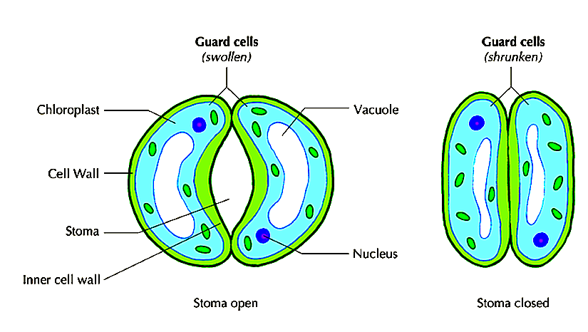
FIGURE 1: STRUCTURE OF GUARD CELL
Mechanism of Opening and Closing of Stomata
Proposed causes of turgor changes in guard cells.
Accumulation of sugar.
Explanation is based on accumulation of potassium
Ions
Process of Gaseous Exchange in Root Stem and Leaves of Aquatic and Terrestrial Plants
Gaseous Exchange in leaves of Terrestrial Plants
Gaseous exchange takes place by diffusion. The structure of the leaf is adapted for gaseous exchange by having intercellular spaces that are filled. These are many and large in the spongy mesophyll.
When stomata are open, carbon (IV) oxide from the atmosphere diffuses into the substomatal air chambers.
From here, it moves into the intercellular space in the spongy mesophyll layer. The CO2 goes into solution when it comes into contact with the cell surface and diffuses into the cytoplasm. A concentration gradient is maintained between the cytoplasm of the cells and the intercellular spaces. CO2 therefore continues to diffuse into the cells. The oxygen produced during photosynthesis moves out of the cells and into the intercellular spaces. From here it moves to the substomatal air chambers and eventually diffuses out of the leaf through the stomata. At night oxygen enters the cells while CO2 moves out. Gaseous exchange in the leaves of aquatic (floating) plants
Transverse section of leaves of an aquatic plant such as Nymphaea differs from that of terrestrial plant. The following are some of the features that can be observed in the leave of an aquatic plant;
Gaseous Exchange through Stems
Terrestrial Plants
Stems of woody plants have narrow openings or slits at intervals called lenticels. They are surrounded by loosely arranged cells where the bark is broken. They have many large air intercellular spaces through which gaseous exchange occurs. Oxygen enters the cells by diffusion while carbon (IV) oxide leaves. Unlike the rest of the bark, lenticels are permeable to gases and water. Aquatic Plant Stems
The water lily, Salvia and Wolfia whose stems remain in water are permeable to air and water.
Oxygen dissolved in the water diffuses through the stem into the cells and carbon (IV) oxide diffuses out into the water. Gaseous Exchange in Roots
Terrestrial Plants
Gaseous exchange occurs in the root hair of young terrestrial plants. Oxygen in the air spaces in the soil dissolves in the film of moisture surrounding soil particles and diffuses into the root hair along a concentration gradient. It diffuses from root hair cells into the cortex where it is used for respiration. Carbon (IV) oxide diffuses in the opposite direction. In older roots of woody plants, gaseous exchange takes place through lenticels. Aquatic Plants Roots of aquatic plants e.g. water lily are permeable to water and gases. Oxygen from the water diffuses into roots along a concentration gradient. Carbon (IV) oxide diffuses out of the roots and into the water. The roots have many small lateral branches to increase the surface area for gaseous exchange. They have air spaces that help the plants to float. Mangroove plants grow in permanently waterlogged soils, muddy beaches and at estuaries. They have roots that project above the ground level. These are known as breathing roots or pneumatophores. These have pores through which gaseous exchange takes place e.g. in Avicenia the tips of the roots have pores. Others have respiratory roots with large air spaces. TOPICAL QUESTIONSThese questions are good for group discussions in and out of a classroom environment they can also be used in a question and answer brainstorming sessions
PRINTABLES AND DOWNLOADABLES
knec topical questions and answers
other mocks/kcse questions and answers
SPONTANIOUS PODCASTS AND VIDEOSPRINT/DOWNLOAD THESE NOTESSPECIFIC OBJECTIVES
By the end of the topic, the learner should be able to:
TOPIC / SUB-TOPIC BREAKDOWN
THE CELL
Introduction
The microscope
The microscope is used to magnify objects.
Magnification
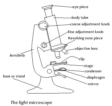
Microscope parts and their functions
To View the Object
Care of a Microscope
Cell Structure as Seen Through the Light Microscope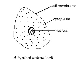
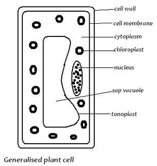
The cell as seen above has the following:
Cell membrane (Plasma membrane):
Cytoplasm:
Vacuole:
Cell wall:
Chloroplasts;
The Electron Microscope (EM)
Cell Structure as Seen Through Electron Microscope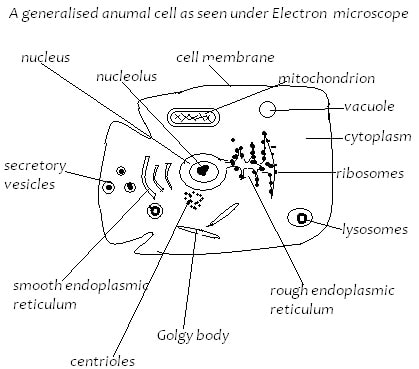
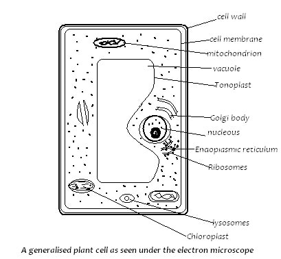
The Plasma Membrane
The Endoplasmic Reticulum (ER)
The Ribosomes
Golgi Bodies
Mitochondria
Lysosomes
The Nucleus
The Chloroplasts
Comparison between animal cell and plant cell
Cell Specialization
Cells are specialized to perform different functions in both plants and animals.
Example;
Animal Tissues
Examples of animal tissues;
Plant Tissues
Example of plant tissues;
Organs
Organ systems
Organism
Practical Activities
Observation and Identification of parts of a light microscope and their functions
Preparation and Observation of Temporary Slides of Plant Cells
Observation of permanent slides of animal cells
Observation and Estimation of Cell Size and Calculation of Magnification of Plant Cells.
These are fats and oils.
Fats are solid at room temperature while oils are liquid. They are made up of carbon, oxygen and hydrogen atoms. The structural units of lipids are fatty acids and glycerol. Fatty acids are made up of hydrocarbon chain molecules with a carboxyl group (-COOH) at one end. In the synthesis of a lipid, three fatty acid molecules combine with one glycerol molecule to form a triglyceride. Three molecules of water are lost in the process. This is a condensation reaction and water is given off. Lipids are hydrolysed e.g. during digestion to fatty acids and glycerol, water is added. Condensation = Glycerol + 3 Fatty hydrolysis Lipid + Water acids 
Properties of Lipids
Proteins
Essential and Non-Essential Amino Acids
Formation of Proteins
Functions of Proteins
As structural materials proteins-
As functional chemical compounds. Examples are hormones and enzymes that act as regulators in the body. Respiratory pigments. Examples are haemoglobin that transports oxygen in the blood and myoglobin that stores up oxygen in muscles. Contractile proteins - make up muscles, i.e. myosin and actin. Proteins combine with other chemical groups to form important substances e.g. mucin in saliva. Source of energy. Proteins are a source of energy in extreme conditions when carbohydrates and fats are not available e.g. in starvation.
Enzymes
Properties of Enzymes
Naming of enzymes
Enzymes are named by adding the suffix -ase to: Name of substrate that they work on e.g.
Factors Affecting Enzyme Action
Temperature
TOPICS
Lesson Objectives
By the end of the topic, the learner should be able to:
Meaning of cell physiology
The term physiology refers to the functions that occur in living organisms.
Cell physiology refers to the process through which substances move across the cell membrane. Several physiological processes take place inside the cell e.g. respiration. Oxygen and glucose required enter the cell while carbon (IV) oxide and water produced leave the cell through the cell membrane. 
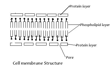
Structure and properties of cell membrane
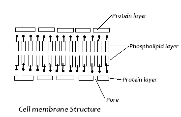
Properties of cell membrane
Permeability
Concentration Gradient
An increase in the concentration of molecules at one region results in a steeper concentration gradient which in turn increases the rate of diffusion. Temperature High temperature increases kinetic energy of molecules. They move faster hence resulting in an increase in rate of diffusion, and vice versa. Size of Molecules or Ions The smaller the size of molecules or ions, the faster their movement hence higher rate of diffusion. Density The denser the molecules or ions diffusing, the slower the rate of diffusion, and vice versa. Medium The medium through which diffusion occurs also affects diffusion of molecules or ions. For example, diffusion of molecules through gas and liquid media is faster than through a solid medium. Distance This refers to the thickness or thinness of surface across which diffusion occurs. Rate of diffusion is faster when the distance is small i.e., thin surface. Surface Area to Volume Ratio The larger the surface area to volume ratio, the faster the rate of diffusion. For example, in small organisms such as Amoeba the surface area to volume ratio, is greater hence faster diffusion than in larger organisms. Factors Affecting DiffusionRole of Diffusion in Living Organisms
Some processes that depend on diffusion include the following:
Osmosis
Factors Affecting Osmosis
Size of solute molecules-
Osmosis' occurs only when solute molecules are too large to pass through a semi-permeable membrane. Concentration Gradient. Osmosis occurs when two solutions of unequal solute concentration are separated by a semi-permeable membrane. Temperature. High temperatures increase movement of water molecules hence influence osmosis. However, too high temperatures denature proteins in cell membrane and osmosis stops. Pressure Increase in pressure affects movement of water molecules. As pressure increases inside a plant cell, osmosis decreases. Roles of Osmosis in Living Organisms
The following processes depend on osmosis in living organisms:
Water Relations in Plant and Animal Cells
Osmotic Pressure
Plasmolysis
Wilting
Water Relations in Plants and AnimalsHaemolysis
Crenation
Active Transport
Factors Affecting Active Transport:
Temperature
Availability of carbohydrates
Metabolic poisons
Role of Active Transport in Living Organisms
Processes requiring active transport:
Practical Activities
1. Experiment to Demonstrate Diffusion
4. Experiment to Demonstrate Turgor and Plasmolysis in Onion Epidermal Cells
CELL PHYSIOLOGY QUESTIONS
1. 1994 Q6 P1
Give a reason for each of the following a) A mature plant cell does not lose its shape even after losing water. b) Xylem vessels do not collapse even when they do not contain water. 2. 1995 Q4 P1 Explain what would happen to red blood cells if they are placed in a concentrated salt solution ( 2 marks) 3. 1995 Q10 P1 An experiment was carried out to investigate the rate of reaction shown below Sucrose →Fructose + Glucose For the products fructose and glucose to be formed, it was found that substance K was to be added and the temperature maintained at 370C. When another substance L was added, the reaction slowed down and eventually stopped. (a) Suggest the identify of substances K and L (2 marks) (b) Other than temperature state three ways by which the rate of reaction could be increased (3 marks) (c) Explain how substance L slowed down the reaction (2 marks) 4. 2000 Q8 P1 Why is oxygen important in the process of active transport in cells? 5. 2004 Q16 P1 a) What is diffusion (2 marks) b) How do the following factors affect the rate of diffusion? i) Diffusion gradient (1 mark) ii) Surface area volume ratio (1mark) iii) Temperature (1mark) c) Outline three roles of active transport in the human body (3 marks) 6. 2005 Q7 P1 State the importance of osmosis in plants. (3 marks) 7. 2009 Q13 P1 (a) Distinguish between diffusion and active transport (2 marks) (b) State one role that is played by osmosis in (1 mark) (i) Plants (ii) Animals 8. 2010 Q7 P1 Distinguish between haemolysis and plasmolysis. (2 marks) FORM 1 BIOLOGY RESOURCES
BIOLOGY TEXTBOOKS 2017/2018 FORM 1
EAST AFRICAN EDUCATIONAL PUBLISHERS Certificate Biology Form 1 ~ Kshs. 458 FOCUS - HIGHFLYER Secondary K.C.S.E Revision Biology ~ Kshs. 465 JOMO KENYATTA FOUNDATION - JYOTI BUNDU - KLB Secondary Biology Book 1 ~ Kshs. 638 LONGHORN Longhorn Secondary Biology Form 1 ~ Kshs. 550 LONGMAN OVERSEAS - LONGMAN KENYA Explore Biology Form 1 ~ Kshs. 618 MALIMU PUBLICATIONS KCSE Revision Series Q & A ~ Kshs. 360 MENTOR PUBLICATIONS - MORAN PUBLISHERS KCSE Golden Tips Revision Series Biology ~ Kshs. 765 Secondary Breakthrough Series Biology Form 1 ~ Kshs. 539 MOTIVATING EDUCATIONAL How to Pass Biology Form 1 & 2 ~ Kshs. 522 MOUNTAIN TOP PUBLISHERS - OXFORD UNIVERSITY PRESS Comprehensive Secondary Biology Form 1 ~ Kshs. 603 Test it & Fix it KCSE Revision Series ~ Kshs. 810 PEZI Principles of Biology Vol.1 3rd Edition ~ Kshs. 590 GOLDEN BELL - SPOTLIGHT PUBLISHERS Spotlight Quick Revision Biology F1 & 2 ~ Kshs. 700 KCSE Mirror Series Biology ~ Kshs. 910 TARGETER PRINTING PRESS - MARIMBA PUBLISHERS LTD - Introduction to Biology NotesTOPIC OBJECTIVES
By the end of the topic, the learner should be able to:
Topics
INTRODUCTION (5 LESSONS)
Introduction to Biology
Biology derived from Greek words
Biology is therefore the study of living things/organisms. 
Branches of Biology
Importance of Biology
Characteristics of Living Things
Life defined through observations of activities carried out by living things;
Nutrition Nutrition is the processes by which food/nutrients are acquired/made and utilized by living organisms. Green plants and certain bacteria make their own food. All other organisms feed on complex organic materials. Respiration This is the breakdown of food to provide energy. The energy released is used for various activities in the organism. Gaseous Exchange Process through which respiratory gases (CO2 & O2) are taken in and out through a respiratory surface. Excretion Excretion is the removal of metabolic wastes from the body. Substances like urea, carbon dioxide (Carbon (IV) oxide). These substances are poisonous if allowed to accumulate in the body. Growth and Development Growth means irreversible change in size. All organisms increase in size that is, they grow. Development is irreversible change in complexity. As they do so, they also become differentiated in form. Reproduction Reproduction is the formation of new individuals of a species to ensure continued existence of a species and growth of its population. Irritability The ability of organisms to detect and respond to changes in the environment. This is of great survival value to the organism. Movement This the progressive change in position from one place to another. Some organisms are sessile (i.e. fixed to the substratum). The majority of plants move only certain parts. Collection and Observation of Organisms
Biology as a practical subject is learnt through humane handling of organisms.
Materials needed for collection of organisms include:-
Observation of Organisms
Presenting the Results of Observations
Organisms are observed and important features noted down: colour, texture hard or soft; if hairy or not. Size is measured or estimated.
Biological DrawingsIt is necessary to draw some of the organisms. In making a biological drawing, magnification (enlargement) is noted. Indicate the magnification of your drawing, i.e. how many times the drawing is larger/smaller than the actual specimen MG=length of drawing/length specimen How to Draw
Collection, Observation and Recording of Organisms
Collection
Plants and animals collected from the environment, near school or within school compound using nets, bottles and gloves. Animals collected include:- arthropods, earthworms and small vertebrates like lizards/chameleons/ rodents. Place in polythene bags and take to the laboratory. Stinging/poisonous insects killed using ether. Other animals are observed live and returned to their natural habitat. Plant specimen collected include: - leaves, flowers and whole plants. Observations are made to show the following:-
The differences between animals and plants collected.
COMPARISON BETWEEN PLANTS AND ANIMALS
Introduction to Biology Questions and AnswersTransport in AnimalsThe Circulatory System Agriculture Form 1 Notes
Large and complex animals have circulatory systems that consist of tubes, a transport fluid and a means of pumping the fluid.
Blood is the transport fluid which contains dissolved substances and cells. The tubes are blood vessels through which dissolved substances are circulated around the body. The heart is the pumping organ which keeps the blood in circulation. The types of circulatory system exist in animals: open and closed.
In an open circulatory system;
The heart pumps blood into vessels which open into body spaces known as haemocoel. Blood comes into contact with tissues. A closed circulatory system; Found in vertebrates and annelids where the blood is confined within blood vessels and does not come into direct contact with tissues. 
Transport in Insects
Mammalian Circulatory System
The heart undergoes contraction (systole) and relaxation (diastole). Systole When the ventricular muscles contract, the cuspid valves (tricuspid and bicuspid) close preventing backflow of blood into auricles. The volume of the ventricles decreases while pressure increases. This forces blood out of the heart to the lungs through semi-lunar valves and pulmonary artery, and to the body tissues via semi-lunar valve and aorta respectively. At the same time the atria are filled with blood. The left ventricle has thicker muscles than the right ventricle, and pumps blood for a longer distance to the tissues.
Diastole
When ventricular muscles relax, the volume of each ventricle increases while pressure decreases. Contractions of atria force the bicuspid and tricuspid valves to open allowing deoxygenated blood from right atrium into right ventricle which oxygenated blood flows from left atrium into the left ventricle. Semi-lunar valves close preventing the backflow of blood into ventricles. The slight contractions of atria force the, blood flow into ventricles. The Heartbeat The heart is capable of contracting and relaxing rhythmically without fatigue due to its special muscles called cardiac muscles. The rhythmic contraction of the heart arise from within the heart muscles without nervous stimulation. The contraction is said to be myogenic. The heartbeat is initiated by the pacemaker or sino-artrio-node (SAN) which is located in the right atrium. The wave of excitation spreads over the walls of atria. It is picked by the artrio-ventricular node which is located at the junction: Of the atria and ventricles, from where the purkinje tissue spreads the wave to the walls of the ventricles. The heart contracts and relaxes rhythmically at an average rate of 72 times per minute. The rate of the heartbeat is increased by the sympathetic nerve, while it is slowed down by the vagus nerve. Heartbeat is also affected by hormones e.g. adrenaline raises the heartbeat. Structure and Function of Arteries, Capillaries and Veins
Arteries
Arteries carry blood away from the heart. They carry oxygenated blood except pulmonary artery which carries deoxygenated blood to the lungs. Arteries have a thick, muscular wall, which has elastic and collagen fibres that resist the pressure of the blood flowing in them. The high pressure is due to the pumping action of the heart. The pressure in the arteries originate from the pumping action of the heart. The pulse or number of times the heart beats per minute can be detected by applying pressure on an artery next to the bone e.g. by placing the finger/thumb on the wrist. The innermost layer of the artery is called endothelium which is smooth. It offers least possible resistance to blood flow. Have a narrow lumen. The aorta forms branches which supply blood to all parts of the body. These arteries divide into arterioles which further divide to form capillaries. Capillaries Capillaries are small vessels whose walls are made of endothelium which is one cell thick. This provides a short distance for exchange of substances. Capillaries penetrate tissues, The lumen is narrow therefore blood flowing in capillaries is under high pressure. Pressure forces water and dissolved substances out of the blood to form tissue fluid. Exchange of substances occurs between cells and tissue fluid. Part of the tissue fluid pass back into capillaries at the venule end. Excess fluid drains into small channels called lymph capillaries which empty their contents into lymphatic vessels. Capillaries join to form larger vessels called venules which in turn join to form veins which transport blood back to the heart. Veins Veins carry deoxygenated blood from the tissues to the heart (except pulmonary vein which carries oxygenated blood from the lungs to the heart). Veins have a wider lumen than arteries. Their walls are thinner than those of arteries. Blood pressure in the veins is low. Forward flow of blood in veins is assisted by contraction of skeletal muscles, hence the need for exercise. Veins have valves along their length to prevent backflow of blood. This ensures that blood flows towards the heart. The way the valves work can be demonstrated on the arm. By pressing on one vein with two fingers, leaving one and pushing blood toward the heart then releasing the latter finger, it can be observed that the part in between is left with the vein not being visible. This is because bleed does not flow back towards the first finger. Diseases and Defects of Circulatory System
Thrombosis
Formation of a clot in the blood vessels is called thrombosis. Coronary thrombosis is the most common. It is caused by blockage of coronary artery which supplies blood to the heart. Blockage may be due to artery becoming fibrous or accumulation of fatty material on the artery walls. Narrow coronary artery results in less blood reaching the heart muscles. A serious blockage can result in heart attack which can be fatal. Heavy intake of fat, alcohol, being overweight and emotional stress can cause coronary thrombosis. A blockage in the brain can lead to a stroke causing paralysis of part of the body, coma or even death. A healthy lifestyle, avoiding a lot of fat in meals and avoiding alcohol can control the disease. Arteriosclerosis This condition results from the inner walls having materials being deposited there or growth of fibrous connective tissue. This leads to thickening of the wall of the artery and loss of elasticity. Normal blood flow is hindered. Arteriosclerosis can lead to thrombosis or hypertension. A person with hypertension which is also called high blood pressure has his/her blood being pumped more forcefully through the narrow vessels. This puts stress on the walls of the heart and arteries. Regular exercise, healthy diet and avoiding smoking can help maintain normal blood pressure. Varicose Veins Superficial veins especially at the back of the legs become swollen and flabby due to some valves failing to function properly. This results to retention of tissue fluid. Regular physical exercise will prevent this condition. Repair of valves through surgery can also be done. Wearing surgical stockings may ease a mild occurrence. Structure and Function of Blood
Composition of Blood
The mammalian blood is made up of a fluid medium called plasma with substances dissolved in it. Cellular components suspended in plasma include;
This is a pale yellow fluid consisting of 90% water. There are dissolved substances which include;
The functions of plasma include: Transport of red blood cells which carry oxygen. Transport dissolved food substances round the body. Transport metabolic wastes like nitrogenous wastes and carbon (IV) oxide in solution about 85% of the carbon (IV) oxide is carried in form of hydrogen carbonates. Transport hormones from sites of production to target organs. Regulation of pH of body fluids. Distributes heat round the body hence regulate body temperature. Erythrocytes (Red Blood Cells) In humans these cells are circular biconcave discs without nuclei. Absence of nucleus leaves room for more haemoglobin to be packed in the cell to enable it to carry more oxygen. Haemoglobin contained in red blood cells is responsible for the transport of oxygen. 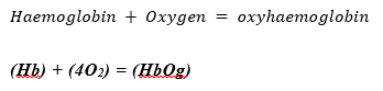
Oxygen is carried in form of oxyhaemoglobin.
Haemoglobin readily picks up oxygen in the lungs where concentration of oxygen is high. In the tissues, the oxyhaemoglobin breaks down (dissociates) easily into haemoglobin and oxygen. Oxygen diffuses out of the red blood cells into the tissues. Haemoglobin is then free to pick up more oxygen molecules. The biconcave shape increases their surface area over which gaseous exchange takes place. Due to their ability, they are able to change their shape to enable themselves squeeze inside the narrow capillaries. There are about five million red blood cells per cubic millimetre of blood. They are made in the bone marrow of the short bones like sternum, ribs and vertebrae. In the embryo they are made in the liver and spleen. Erythrocytes have a life span of about three to four months after which they are destroyed in the liver and spleen. Also in the red blood cells is carbonic anhydrase which assists in the transport of carbon (IV) oxide. Leucocytes (White Blood Cells) These white blood cells have a nucleus. They are divided into two:
Neutrophils form 70% of the granulocytes. Others are eosinophils and basophils. About 24% agronulocytes are called lymphocytes, while 4% agranulocytes are monocytes. The leucocytes are capable of amoebic movement. They squeeze between the cells of the capillary wall to enter the intercellular spaces. They engulf and digest disease causing organisms (pathogens) by phagocytosis. Some white blood cells may die in the process of phagocytosis. The dead phagocytes, dead organisms and damaged tissues form pus. Lymphocytes produce antibodies which inactivate antigens. Antibodies include: Antitoxins which neutralise toxins. Agglutinins cause bacteria to clump together and they die. Lysins digest cell membranes of microorganisms. Opsonins adhere to outer walls of microorganisms making it easier for phagocytes to ingest them. Lymphocytes' are made in the thymus gland and lymph nodes. There are about 7,000 leucocytes per cubic millimetre of blood. Platelets (Thrombocytes) Platelets are small irregularly shaped cells formed from large bone marrow cells called megakaryocytes. There are about 250,000 platelets per cubic millimetre of blood. They initiate the process of blood clotting. The process of clotting involves a series of complex reactions whereby fibrinogen is converted into a fibrin clot. When blood vessels are injured platelets are exposed to air and they release thromboplastin which initiates the blood clotting process. Thromboplastin neutralises heparin the anti-clotting factor in blood and activates prothrombin to thrombin. The process requires calcium ions and vitamin K. Thrombin activates the conversion of fibrinogen to fibrin which forms a meshwork of fibres on the cut surface to trap red blood cells to form a clot. The clot forms a scab that stops bleeding and protects the damaged tissues from entry of micro-organisms. Blood clotting reduces loss of blood when blood vessels are injured. Excessive loss of blood leads to anaemia and dehydration. Mineral salts lost in blood leads to osmotic imbalance in the body. This can be corrected through blood transfusion and intravenous fluid. ABO Blood Groups There are four types of blood groups in human beings: A, B, AB and O. These are based on types of proteins on the cell membrane of red blood cells. There are two types of proteins denoted by the letters A and B which are antigens. In the plasma are antibodies specific to these antigens denoted as a and b. A person of blood group A has A antigens on the red blood cells and b antibodies in plasma. A person of blood group B has B antigens on red blood cells and a antibodies in plasma. A person of blood group AB has A and B antigens on red blood cells and no antibodies in plasma. A person of blood group a has no antigens on red blood cells and a and b antibodies in plasma. 
Blood Transfusion
Blood transfusion is the transfer of blood from a donor to the circulatory system of the recipient.
A recipient will receive blood from a donor if the recipient has no corresponding antibodies to the donor's antigens. If the donor's blood and the recipient's blood are not compatible, agglutination occurs whereby red blood cells clump together. Blood typing A person of blood group 0 can donate blood to a person of any other blood group. A person of blood group 0 is called a universal donor. A person of blood group AB can receive blood from any other group. A person with blood group AB is called a universal recipient. A person of blood group A can only donate blood to another person with blood group A or a person with blood group AB. A person of blood group B can only donate blood to somebody with blood group B or a person with blood group AB. A person with blood group AB can only donate blood to a person with blood groupAB. Blood screening has become a very important step in controlling HIV/AIDS. It is therefore important to properly screen blood before any transfusion is done. Rhesus factor The Rhesus factor is present in individuals with the Rhesus antigen in their red blood cells. Such individuals are said to be Rhesus positive (Rh+), while those without the antigen are Rhesus negative (Rh-). If blood from an Rh+ individual is introduced into a person who is Rh- , the latter develops antibodies against the Rhesus factor. There may not be any reaction after this transfusion. However a subsequent transfusion with Rh+ blood causes a severe reaction, and agglutination occurs i.e. clumping of red blood cells. The clump can block the flow of blood, and cause death. Erythroblastosis foetalis (haemolytic disease of the newborn) results when an Rh- mother carries an Rh+ foetus. This arises when the father is Rh+. During the latter stage of pregnancy, fragments of Rhesus positive red blood cells of the foetus may enter mother's circulation. These cause the mother to produce Rhesus antibodies which can pass across the placenta to the foetus and destroy foetal red blood cells. During the first pregnancy, enough antibodies are not formed to affect the foetus. Subsequent pregnancies result in rapid production of Rhesus antibodies by the mother. These destroy the red blood cells of the foetus, the condition called haemolytic disease of the newborn. The baby is born anaemic and with yellow eyes (jaundiced). The condition can be corrected by a complete replacement of baby's blood with safe healthy blood. Lymphatic System The lymphatic system consists of lymph vessels. Lymph vessels have valves to ensure unidirectional movement of lymph. Lymph is excess tissue fluid i.e. blood minus blood cells and plasma proteins. Flow of lymph is assisted by breathing and muscular contractions. Swellings called lymph glands occur at certain points along the lymph vessels. Lymph glands are oval bodies consisting of connective tissues and lymph spaces. The lymph spaces contain lymphocytes which are phagocytic. Lymph has the same composition as blood except that it does not contain red blood cells and plasma proteins. Lymph is excess tissue fluid. Excess tissue fluid is drained into lymph vessels by hydrostatic pressure. The lymph vessels unite to form major lymphatic system. The main lymph vessels empty the contents into sub-clavian veins which take it to the heart. Immune Responses Immune response is the production of antibodies in response to antigens. An antigen is any foreign material or organism that is introduced into the body and causes the production of antibodies. Antigens are protein in nature. An antibody is a protein whose structure is complementary to the antigen. This means that a specific antibody deals with a specific antigen to make it harmless. When harmful organisms or proteins invade the body, lymphocytes produce complementary antibodies, while bone marrow and thymus gland produce more phagocytes and lymphocytes respectively. Types of Immunity There are two types of immunity; natural and artificial. Natural Immunity is also called innate immunity. It is inherited from parent to offspring. Artificial Immunity can be natural or induced. When attacked by diseases like chicken pox, measles and mumps, those who recover from these diseases develop resistance to any subsequent infections of the same diseases. This is natural acquired immunity. Artificial Acquired Immunity: When attenuated (weakened) or dead microorganisms are introduced into a healthy person. The lymphocytes synthesis the antibodies which are released into the lymph and eventually reach the blood. The antibodies destroy the invading organisms. The body retains 'memory' of the structure of antigen. Rapid response is ensured in subsequent infections. Vaccines generally contain attenuated disease causing organisms.
Artificial Passive Acquired Immunity:
Serum containing antibodies is obtained from another organism, and confers immunity for a short duration. Such immunity is said to be passive because the body is not activated to produce the antibodies. Importance of Vaccination A vaccine is made of attenuated, dead or non-virulent micro-organism that stimulate cells in the immune system to recognise and attack disease causing agent through production of antibodies. Vaccination protects individuals from infections of many diseases like smallpox, tuberculosis and poliomyelitis. Diseases like smallpox, tuberculosis and tetanus were killer diseases but this is no longer the case. Diphtheria Pertussis Tetanus (DPT) vaccine protects children against diphtheria, whooping cough and tetanus. Bacille Calmette Guerin (BCG) vaccine is injected at birth to children to protect them against tuberculosis. Measles used to be a killer disease but today, a vaccine injected into children at the age of rune months prevents it. At birth children are given an inoculation through the mouth of the poliomyelitis vaccine. Allergic Reactions An allergy is a hypersensitive reaction to an antigen by the body. The antibody reacts with the antigen violently. People with allergies are oversensitive to foreign materials like dust, pollen grains, some foods, some drugs and some air pollutants. Allergic reactions lead to production of histamine by the body. Histamine causes swelling and pain. Allergic reactions can be controlled by avoiding the allergen and administration of anti-histamine drugs. transport in animals videos.
|
|||||||||||||||||||||||||||||||||||||||||||||||||||||||||||||||||||||||||||||||||||||||||||||||||||||||||||||||||||

Heterotrophism
Meaning and Types of Heterotrophism
- This is a mode of nutrition whereby organisms feed on complex organic matter from other plants or animals.
- All animals are heterotrophs.
- Their mode of feeding is also said to be holozoic to distinguish it from other special types of heterotrophic nutrition namely:
- Saprophytism
- Parasitism
- Saprophytism/saprotrophysim- occurs in most fungi and some forms of bacteria.
- Saprophytes feed on dead organic matter and cause its decomposition or decay.
- Parasitism is a mode of feeding whereby one organism called the parasite feeds on or lives in another organism called the host and harms it.
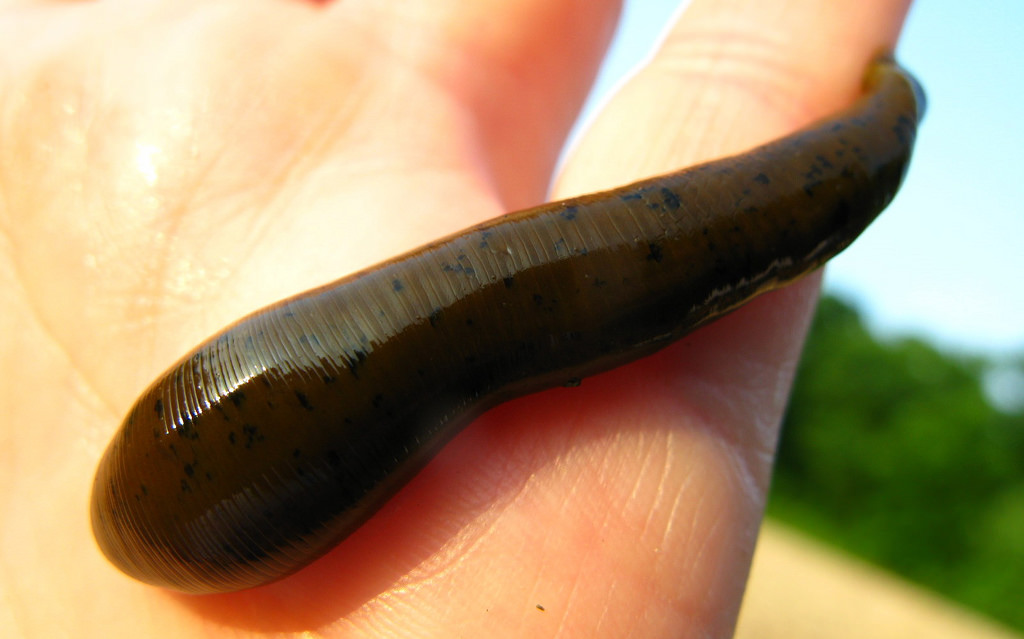
Leech
Modes of Feeding in Animals
- Animals have developed various structures to capture and ingest food.
- The type of structures present depend on the method of feeding and the type of food.
- Carnivorous animals feed on whole animals or portions of their flesh.
- Herbivorous animals feed on plant material.
- Omnivorous animals feed on both plants and animal materials.
Feeding in Mammals
- The jaws and teeth of mammals are modified according to the type of food eaten.
- Mammals have different kinds of teeth.
- Each type of teeth has a particular role to play in the feeding process.
Feeding in Mammals
- The jaws and teeth of mammals are modified according to the type of food eaten.
- Mammals have different kinds of teeth.
- Each type of teeth has a particular role to play in the feeding process.
Feeding in Mammals
- The jaws and teeth of mammals are modified according to the type of food eaten.
- Mammals have different kinds of teeth.
- Each type of teeth has a particular role to play in the feeding process.
- This condition is described as heterodont.
- The teeth of reptiles and amphibians are all similar in shape and carry out the same function.
- They are said to be homodont.
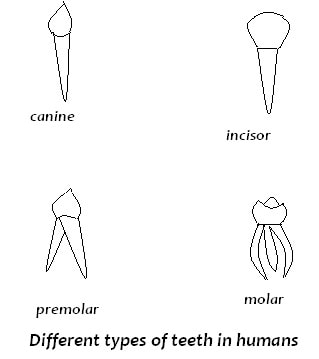
Types of Mammalian Teeth
- Mammals have four kinds of teeth.
- The incisors are found at the front of the jaw.
- They are sharp-edged and are used for biting.
- The canines are located at the sides of the jaw.
- They are pointed and are used for tearing and piercing.
- The premolars are next to the canines and the molars are at the back of the jaw.
- Both premolars and molars are used for crushing and grinding.
- Teeth are replaced only once in a lifetime.
- The first set is the milk or deciduous teeth.
- These are replaced by the second set or the permanent teeth.
- Dentition refers to the type of teeth, the number and their arrangement in the jaw.
- A dental formula shows the type and number of teeth in each half of the jaw.
- The number of teeth in half of the upper jaw is represented above a line and those on the lower jaw below the line.
- The first letter of each type of teeth is used in the formula i.e. i = incisors, c = canines, pm = premolars and m = molars.
- The total number is obtained by multiplying by two (for the two halves of each jaw).
Adaptation of Teeth to Feeding
In general, incisors are for cutting, canines for tearing while premolars and molars are for grinding.
However, specific modifications are observed in different mammals as an adaptation to the type of food they eat.
Teeth of Herbivores
In general, incisors are for cutting, canines for tearing while premolars and molars are for grinding.
However, specific modifications are observed in different mammals as an adaptation to the type of food they eat.
Teeth of Herbivores
- Incisors are long and flat with a sharp chisel like edge for cutting.
- The enamel coating is thicker in front than at the back so that as the tooth wears out, a sharp edge is maintained.
- Canines are reduced or absent.
- If absent, the space left is called the diastema.
- The diastema allows the tongue to hold food and push it to the grinding teeth at the back of the mouth.
- These are transversely ridged.
- The ridges on the upper teeth fit into grooves on the lower ones.
- This gives a sideways grinding surface.
- The teeth of herbivores have open roots i.e., wide opening into the pulp cavity.
- This ensures a continued adequate supply of food and oxygen to the tooth.
- In some herbivores, such as rabbits and elephants, the incisors continue to grow throughout life.
Teeth of Carnivores
- Incisors are reduced in size and pointed.
- They are well suited for grasping food and holding prey.
- Canines are long, pointed and curved.
- They are used for piercing and tearing flesh as well as for attack and defence.
- Premolars and molars: In general, they are long and longitudinally ridged to increase surface area for crushing.
- Carnassial Teeth: These are the last premolars on the upper jaw and the first molars on the lower one.
- They are enlarged for cutting flesh.
- They act as a pair of shears.
- They also crush bones.
- The teeth of carnivores have closed roots i.e., only a very small opening of the pulp cavity to allow food and oxygen to keep teeth alive.
- Once broken, no re-growth can take place.
- Incisors have a wide surface for cutting.
- Canines are bluntly pointed for tearing.
- Premolars and molars have cusps for crushing and grinding.
- The premolars have two blunt cusps while the molars have three to four.
Internal Structure of tooth
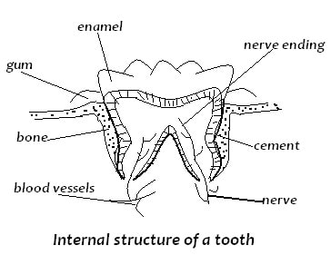
The tooth consists of two main parts:
Crown: The portion above the gum; it is covered by the enamel.
Root: The portion below the gum; it is covered by the cement.
The tooth has two roots.
Neck: Is the region at the same level with the gum.
Cement: Fixes the tooth firmly to the jaw bone.
Crown: The portion above the gum; it is covered by the enamel.
Root: The portion below the gum; it is covered by the cement.
The tooth has two roots.
Neck: Is the region at the same level with the gum.
- It forms the junction between the crown and the root.
- It is covered by enamel. Incisors and canines have one root only.
- Premolars have one or two roots while molars have two to three roots each.
- Internally, the bulk of the tooth is made up of dentine which consists of living cells and extends to the root.
- It is composed of calcium salts, collagen and water.
- It is harder than bone but wears out with use.
- This is why it is covered by enamel which is the hardest substance in a mammal's body.
Cement: Fixes the tooth firmly to the jaw bone.
Common Dental Diseases
Dental Carries
Dental carries are the holes or cavities that are formed as acid corrodes enamel and eventually the dentine.
Causes
Treatment depends on the extent of the dental caries:
Extraction of Tooth.
Filling
This involves replacing the dentine with amalgam, a mixture of hard elements e.g. silver and tin.
Root Canal Treatment
This involves surgery and reconstruction.
It saves severely damaged teeth.
The nerves in the root canal are surgically severed.
The tooth is cleaned and filled up with amalgam.
Periodontal Diseases
Dental carries are the holes or cavities that are formed as acid corrodes enamel and eventually the dentine.
Causes
- This is caused by bacteria acting on the food left between teeth and on the cusp.
- Acids are formed that eventually corrode the enamel.
- The pulp cavity is eventually reached.
- A lot of pain is experienced then.
- The bacteria then infect the pulp cavity and the whole tooth decays.
Treatment depends on the extent of the dental caries:
Extraction of Tooth.
Filling
This involves replacing the dentine with amalgam, a mixture of hard elements e.g. silver and tin.
Root Canal Treatment
This involves surgery and reconstruction.
It saves severely damaged teeth.
The nerves in the root canal are surgically severed.
The tooth is cleaned and filled up with amalgam.
Periodontal Diseases
- These are diseases of the gum.
- The gum becomes inflamed, and starts bleeding.
- Progression of the disease leads to infection of the fibres in the periodontal membranes and the tooth becomes loose.
- This condition is known as pyorrhoea.
- The diseases are caused by poor cleaning of the teeth.
- The accumulation of food particles leading to formation of plaque, lack of adequate vitamin A and C in the diet.
- Nutrition - by taking adequate balanced diet rich in vitamins A and C.
- Antibiotics are used to kill bacteria.
- Anti-inflammatory drugs are given.
- Antiseptic is prescribed to use in cleaning the mouth daily to prevent further proliferation of bacteria.
- The plaque is removed-drilled away - a procedure known as scaling.
Care of Teeth
In order to maintain healthy teeth the following points should be observed:
- A proper diet that includes calcium and vitamins, particularly vitamin D is essential.
- The diet should also contain very small quantities of fluorine to strengthen the enamel.
- Large quantities of fluorine are harmful.
- The enamel becomes brown, a condition known as dental fluorosis.
- Chewing of hard fibrous foods like carrots and sugar cane to strengthen and cleanse the teeth.
- Proper use of teeth e.g. not using teeth to open bottles and cut thread.
- Regular and thorough brushing of teeth after meals.
- Dental floss can be used to clean between the teeth.
- Not eating sweets and sugary foods between meals.
- Regular visits to the dentist for checkups.
- Washing the mouth with strong salt solution or with any other mouth wash with antiseptic properties.
Digestive System and Digestion in Humans
Organs that are involved with feeding in humans constitute the digestive system.
Digestive System and Associated Glands
Organs that are involved with feeding in humans constitute the digestive system.
Digestive System and Associated Glands
- Human digestive system starts at the mouth and ends at the anus.
- This is the alimentary canal.
- Digestion takes place inside the lumen of the alimentary canal.
- The epithelial wall that faces the lumen has mucus glands (goblet cells).
- These secrete mucus that lubricate food and prevent the wall from being digested by digestive enzymes.
- Present at specific regions are glands that secrete digestive enzymes.
- The liver and pancreas are organs that are closely associated with the alimentary canal.
- Their secretions get into the lumen and assist in digestions.
- Mouth.
- Oesophagus.
- Stomach.
- Small intestines - consist of duodenum, the first part next to the stomach, ileum - the last part that ends up in a vestigial caecum and appendix which are nonfunctional.
- Large intestines consist of: colon and rectum that ends in the anus.
Ingestion, Digestion and Absorption
- Feeding in humans involves the following processes:
- Ingestion: This is the introduction of the food into the mouth.
- Digestion: This is the mechanical and chemical breakdown of the food into simpler, soluble and absorbable units.
- Absorption: Taking into blood the digested products.
- Assimilation: Use of food in body cells.
- Mechanical breakdown of the food takes place with the help of the teeth.
- Chemical digestion involves enzymes.
- In the mouth, both mechanical and chemical digestion takes place.
- Food is mixed with saliva and is broken into smaller particles by the action of teeth.
- Saliva contains the enzyme amylase.
- It also contains water and mucus which lubricate and soften food in order to make swallowing easy.
- Saliva is slightly alkaline and thus provides a suitable pH for amylase to act on cooked starch, changing it to maltose.
- The food is then swallowed in the form of semisolid balls known as boluses.
- Each bolus moves down the oesophagus by a process known as peristalsis.
- Circular and longitudinal muscles along the wall of the alimentary canal contract and relax pushing the food along.
- In the stomach, the food is mixed with gastric juice secreted by gastric glands in the stomach wall.
- Gastric juice contains pepsin, rennin and hydrochloric acid.
- The acid provides a low pH of 1.5-2.0 suitable for the action of pepsin.
- Pepsin breaks down protein into peptides.
- Rennin coagulates the milk protein casein.
- The stomach wall has strong circular and longitudinal muscles whose contraction mixes the food with digestive juices in the stomach.
Digestion in the Duodenum
- In the duodenum the food is mixed with bile and pancreatic juice.
- Bile contains bile salts and bile pigments.
- The salts emulsify fats, thus providing a large surface area for action of lipase.
- Pancreatic juice contains three enzymes:
- Trypsin which breaks down proteins into peptides and amino acids,
- Amylase which breaks down starch into maltose, and
- Lipase which breaks down lipids into fatty acids and glycerol.
- These enzymes act best in an alkaline medium which is provided for by the bile.
- Epithelial cells in ileum secrete intestinal juice, also known as succus entericus.
- This contains enzymes which complete the digestion of protein into amino acids, carbohydrates into monosaccharides and lipids into fatty acids and glycerol.
- This is the diffusion of the products of digestion into the blood of the animal.
- It takes place mainly in the small intestines though alcohol and some glucose are absorbed in the stomach.
- It is highly coiled.
- The coiling ensures that food moves along slowly to allow time for its digestion and absorption.
- It is long to provide a large surface area for absorption.
- The epithelium has many finger-like projections called villi (singular villus).
- They greatly increase the surface area for absorption.
- Villi have microvilli that further increase the surface area for absorption.
- The wall of villi has thin epithelial lining to facilitate fast diffusion of products of digestion.
- Has numerous blood vessels for transport of the end products of digestion.
- Has lacteal vessels; for absorption of fatty acids and glycerol and transport of lipids.
- Glucose and other monosaccharides as well as amino acids are absorbed through the villi epithelium and directly into the blood capillaries.
- First they are carried to the liver through the hepatic portal vein, then taken to all organs via circulatory system.
Absorption of Fatty Acids and Glycerol
- Fatty acids and glycerol diffuse through the epithelial cells of villi and into the lacteal.
- When inside the villi epithelial cells, the fatty acids combine with glycerol to make tiny fat droplets which give the lacteal a milky appearance.
- The lacteals join the main lymph vessel that empties its contents into the bloodstream in the thoracic region.
- Once inside the blood, the lipid droplets are hydrolysed to fatty acids and glycerol.
- Vitamins and mineral salts are absorbed into the blood capillaries in' the villi. Water is mainly absorbed in the colon.
- As a result the undigested food is in a semi-solid form (faeces) when it reaches the rectum.
- Egestion: This is removal of undigested or indigestible material from the body. Faeces are temporarily stored in the rectum then voided through the anus. Opening of the anus is controlled by sphincter muscles
- Assimilation: This is the incorporation of the food into the cells where it is used for various chemical processes.
- Carbohydrates are used to provide energy for the body.
- Excess glucose is converted to glycogen and stored in the liver and muscles.
- Some of the excess carbohydrates are also converted into fat in the liver and stored in the adipose tissue' (fat storage tissue), in the mesenteries and in the connective tissue under the skin, around the heart and other internal organs.
- Amino acids are used to build new cells and repair worn out ones.
- They are also used for the synthesis of protein compounds.
- Excess amino acids are deaminated in the liver.
- Urea is formed from the nitrogen part.
- The remaining carbohydrate portion is used for energy or it is converted to glycogen or fat and stored.
- Fats are primarily stored in the fat storage tissues.
- When carbohydrates intake is low in the body, fats are oxidised to provide energy.
- They are also used as structural materials e.g. phospholipids in cell membrane. They act as cushion, protecting delicate organs like the heart.
- Stored fats under the skin act as heat insulators.
Summary of digestion in humans
Importance of Vitamins, Mineral Salts, Roughage and Water in Human Nutrition
Vitamins
- These are organic compounds that are essential for proper growth, development and functioning of the body.
- Vitamins are required in very small quantities.
- They are not stored and must be included in the diet.
- Vitamins Band C are soluble in water, the rest are soluble in fat.
- Various vitamins are used in different ways.
- Mineral ions are needed in the human body.
- Some are needed in small amounts while others are needed in very small amounts (trace).
- All are vital to human health.
- Nevertheless, their absence results in noticeable malfunction of the body processes.
- Water is a constituent of blood and intercellular fluid.
- It is also a constituent of cytoplasm.
- Water makes up to 60-70% of total fresh weight in humans.
- No life can exist without water.
- Acts as a medium in which chemical reactions in the body takes place.
- Acts as a solvent and it is used to transport materials within the body.
- Acts as a coolant due to its high latent heat of vaporisation.
- Hence, evaporation of sweat lowers body temperature.
- Takes part in chemical reactions i.e. hydrolysis.
Vitamins, sources, uses and the deficiency disease resulting from their absence in diet
Roughage
A diet is balanced when it contains all the body's nutrient requirements and in the right amounts or proportions.
A balanced diet should contain the following:
This is faulty or bad feeding where the intake of either less or more than the required amount of food or total lack of some food components.
Deficiency Diseases
Deficiency diseases result from prolonged absence of certain components in the diet.
Examples are:
Marasmus:
Lack of enough food results in thin arms and legs, severe loss of fluid, general body wasting, sunken eyes.
Kwashiorkor
Lack of protein in the diet of children. The symptoms of kwashiorkor include wasting of the body, red thin hair, swollen abdomen and scaly skin.
Other deficiency diseases are due to lack of accessory food factors (vitamins and mineral salts.). Such diseases include rickets, goitre and anaemia.
Treatment of these deficiency diseases is by supplying the patient with the component missing in the diet.
- Roughage is dietary fibre and it consists mainly of cellulose.
- It adds bulk to the food and provides grip for the gut muscles to enhance peristalsis.
- Roughage does not provide any nutritional value because humans and all animals not produce cellulase enzyme to digest cellulose.
- In herbivores symbiotic bacteria in the gut produce cellulase that digests cellulose.
- Age: Infants, for instance, need a greater proportion of protein than adults.
- Sex: males generally require more carbohydrates than females.
- The requirements of specific nutrients for females depends on the stage of development in the life cycle.
- Adolescent girls require more iron in their diet; expectant and nursing mothers require a lot of proteins and mineral salts.
- State of Health: A sick individual requires more of certain nutrients e.g. proteins, than a healthy one.
- Occupation: An office worker needs less nutrients than a manual worker.
A diet is balanced when it contains all the body's nutrient requirements and in the right amounts or proportions.
A balanced diet should contain the following:
- Carbohydrates
- Proteins
- Lipids
- Vitamins
- Mineral Salts
- Water
- Dietary fibre or roughage
This is faulty or bad feeding where the intake of either less or more than the required amount of food or total lack of some food components.
Deficiency Diseases
Deficiency diseases result from prolonged absence of certain components in the diet.
Examples are:
Marasmus:
Lack of enough food results in thin arms and legs, severe loss of fluid, general body wasting, sunken eyes.
Kwashiorkor
Lack of protein in the diet of children. The symptoms of kwashiorkor include wasting of the body, red thin hair, swollen abdomen and scaly skin.
Other deficiency diseases are due to lack of accessory food factors (vitamins and mineral salts.). Such diseases include rickets, goitre and anaemia.
Treatment of these deficiency diseases is by supplying the patient with the component missing in the diet.
Practical Activities
- Experiments to show that Carbon (IV) Oxide is necessary for Photosynthesis
- Experiment to Show Effect of Light on Photosynthesis
- Experiment to Show the Effect of Chlorophyll on Photosynthesis
- Experiment To Observe Stomata Distribution in Different Leaves
- Test for Reducing Sugar
- Test for non-reducing sugar
- Test for Lipids;
- Test for Proteins -Biuret Test
- Experiment To Investigate Presence of Enzyme in Living Tissue
- Dissection of a Rabbit to show the Digestive System
TOPIC OBJECTIVES
By the end of the topic, the learner should be able to:
- use the magnifying lens to observe the external features of plants and animals
- record observations of the main external characteristics of living organisms, preserved specimens and photographs
- state the necessity and significance of classification
- name the major units of classification
- state the application of Binomial nomenclature in naming organisms.
topics / sub-topics breakdown
- Review the use of magnifying lens
- External features of plants and animals
- Necessity and significance of classification
- Major units of classification: (naming)
- Kingdoms
- Monera
- protoctista
- fungi
- plantae
- animalia (At least one example of each)
- For kingdom plantae and animalia, cover phylum/division, class, order, family, genus and species. Show relationship between the taxonomic units (Give at least one example of each taxon)
- Kingdoms
- Discussion on Binomial nomenclature
- Practical activities
- Use of collecting nets, cutting instruments and hand lens.
- Collection and detailed observation of:
- small animals e.g. insects
- plants - rhizoids, root systems (taproot, fibrous and adventitious), stems and leaves
- Collection and detailed observation of:
Classification I
Introduction
Classification is putting organisms into groups.
Classification is based on the study of external characteristics of organisms.
It involves detailed observation of structure and functions of organisms.
Organisms with similar characteristics are put in one group.
Differences in structure are used to distinguish one group from another.
The magnifying lens is an instrument that assists in the observation of fine structure e.g. hairs by enlarging them.
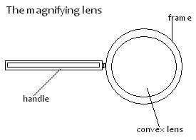
Using a Magnifying Lens
A specimen is placed on the bench or held by hand,
Then the magnifying lens is moved towards the eye until the object is dearly focused and an enlarged image is seen.
The magnification can be worked out as follows:
Note: magnification has no units.
Then the magnifying lens is moved towards the eye until the object is dearly focused and an enlarged image is seen.
The magnification can be worked out as follows:
Note: magnification has no units.
Necessity/need for Classification
To be able to identify organisms into their taxonomic groups.
To enable easier and systematic study of organisms.
To show evolutionary relationships in organisms.
To enable easier and systematic study of organisms.
To show evolutionary relationships in organisms.
Major Units of Classification (Taxonomic Groups)
Taxonomy is the study of the characteristics of organisms for the purpose of classifying them.
The groups are Taxa (singular Taxon).
The taxonomic groups include:
The groups are Taxa (singular Taxon).
The taxonomic groups include:
- Species: This is the smallest unit of classification. Organisms of the same species resemble each other. The number of chromosomes in their cells is the same. Members of a species interbreed to produce fertile offspring.
- Genus (plural genera): A genus is made up of a number of species that share several characteristics. Members of a genus cannot interbreed and if they do, the offspring are infertile.
- Family: A family is made up of a number of genera that share several characteristics.
- Order: A number of families with common characteristics make an order.
- Class: Orders that share a number of characteristics make up a class.
- Phylum/Division: A number of classes with similar characteristics make up a phylum (plural phyla) in animals. In plants this is called a division.
- Kingdom: This is made up of several phyla (in animals) or divisions (in plants). It is the largest taxonomic unit in classification.
Kingdoms
Living organisms are classified into five kingdoms namely;
- Monera,
- Protoctista,
- Fungi,
- Plantae
- Animalia.
Kingdom Fungi
Some are unicellular while others are multicellular.
They have no chlorophyll.
Most are saprophytic e.g. yeasts, moulds and mushrooms.
A few are parasitic e.g. Puccinia graminae.
They have no chlorophyll.
Most are saprophytic e.g. yeasts, moulds and mushrooms.
A few are parasitic e.g. Puccinia graminae.
Kingdom Monera (Prokaryota)
These are very small unicellular organisms.
They lack a nuclear membrane
do not have any bound membrane organelles.
Hence the name Prokaryota.
They are mainly bacteria, e.g. Vibrio cholerae.
They lack a nuclear membrane
do not have any bound membrane organelles.
Hence the name Prokaryota.
They are mainly bacteria, e.g. Vibrio cholerae.
Kingdom Protoctista
They are unicellular organisms.
Their nucleus and organelles are surrounded by membranes (eukaryotic).
They include algae, slime moulds - fungi-like and protozoa
Their nucleus and organelles are surrounded by membranes (eukaryotic).
They include algae, slime moulds - fungi-like and protozoa
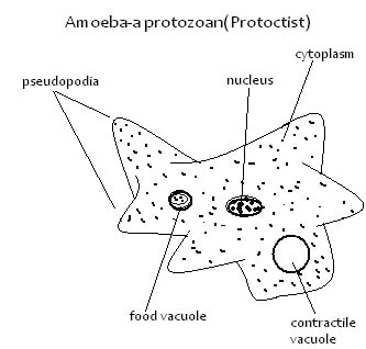
Kingdom Plantae
They are all multicellular.
They contain chlorophyll and are all autotrophic.
They include; Bryophyta (moss plant), Pteridophyta (ferns) and Spermatophyta (seed bearing plants).
They contain chlorophyll and are all autotrophic.
They include; Bryophyta (moss plant), Pteridophyta (ferns) and Spermatophyta (seed bearing plants).
Kingdom Animalia
These are all multicellular and heterotrophic.
Examples are annelida (earthworms), mollusca (snails),athropoda, chordata .
Example of Arthropods are ticks, butterflies.
Members of Chordata are fish, frogs and humans.
Examples are annelida (earthworms), mollusca (snails),athropoda, chordata .
Example of Arthropods are ticks, butterflies.
Members of Chordata are fish, frogs and humans.
External Features of Organisms
In plants we should look for:-
Spore capsule and rhizoids in moss plants.
Sori and fronds in ferns.
Stem, leaves, roots, flowers, fruits and seeds in plants.
In animals, some important features to look for are:
Segmentation, presence of limbs and, number of body parts, presence and number of antennae. These are found in phylum arthropoda:
Visceral clefts, notochord, nerve tube, fur or hair, scales, fins, mammary glands, feathers and wings.
These are found in chordata.
Binomial Nomenclature
Organisms are known by their local names.
Scientists use scientific names to be able to communicate easily among themselves.
This method of naming uses two names, and is called Binomial nomenclature.
The first name is the name of the genus: (generic name) which starts with a capital letter.
The second name is the name of the species (specific name) which starts with a small letter.
The two names are underlined or written in italics.
Man belongs to the genus Homo, and the species, sapiens.
The scientific name of man is therefore Homo sapiens.
Maize belongs to the genus Zea, and the species mays.
The scientific name of maize is Zea mays.
Practical Activities
Use of Collecting Nets, Cutting Instruments and Hand Lens.
Forceps are used to collect crawling and slow moving animals.
Sweep nets are used to catch flying insects.
Cutting instrument like scapel is used to cut specimen e.g. making sections.
Hand lens is used to magnify small plants and animals.
Drawing of the magnified organism are made and the linear magnification of each calculated.
Collection and Detailed Observation of Small Plants and Animals
E.g. moss, ferns, bean.
Look for the following:
Moss plants: Rhizoids and spore capsules.
Fern plants: Rhizomes with adventitious roots; large leaves (fronds) with Sori (clusters of sporangia).
Seed plants: Tree/shrub (woody) or non-woody (herbs) e.g. bean.
Root system - fibrous, adventitious and tap root.
Stem - position and length of internodes.
Type of leaves - simple or compound; arranged as alternate, opposite or whorled.
Flower - colour, number of parts, size and relative position of each:
Fruits - fleshy or dry; edible or not edible.
Seeds - monocotyledonous or dicotyledonous.
Small animals e.g. earthworms, tick, grasshopper, butterfly, beetles.
Observe these animals to see:
- Number of legs.
- Presence or absence of wings.
- Number of antennae.
- Body covering.
- Body parts.
|
Vertical Divider
|
FORM 1ARRANGED ACCORDING TO TOPICS
FORM 2
FORM 3
FORM 4
|
||||||||||||||||||||||||||
Archives
January 2024
November 2023
October 2023
September 2023
March 2023
January 2023
October 2022
September 2021
June 2021
May 2021
Categories
All
CELL PHYSIOLOGY
Classification 1
CLASSIFICATION II
ECOLOGY
EVOLUTION
EXCRETION AND HOMEOSTASIS
FORM 1
FORM 2
FORM 3
FORM 4
Gaseous Exchange In Animals
GASEOUS EXCHANGE IN PLANTS AND ANIMALS
GROWTH & DEVELOPMENT
HUMAN DISEASES
INTRODUCTION TO BIOLOGY
Lipids
MEASUREMENT OF GROWTH
NUTRITION IN ANIMALS
NUTRITION IN PLANTS AND ANIMALS
PDF NOTES
PRACTICAL ACTIVITIES
REPRODUCTION IN PLANTS AND ANIMALS
RESPIRATION
THE CELL
Transport In Animals
TRANSPORT IN PLANTS AND ANIMALS
 RSS Feed
Can't find what you are looking for? Don't worry, Use the Search Box Below.
|
Primary Resources
College Resources
|
Secondary Resources
|
Contact Us
Manyam Franchise
P.O Box 1189 - 40200 Kisii Tel: 0728 450 424 Tel: 0738 619 279 E-mail - sales@manyamfranchise.com |